Cysta (lékařství)
Cysta (z lat. cista – schránka, řeč. kystis – bublina, měchýř) je dutý, patologický útvar, ohraničený od okolní tkáně vlastní, často ale atrofovanou, epiteliální výstelkou. Příčiny vzniku cysty jsou různé, nejčastěji vzniká po uzávěru žlázového vývodu, příčinou může být také jeho rozšíření. Některé cysty jsou důsledkem vývojové anomálie nebo nádorového původu. Zvláštním druhem cyst jsou cysty parazitární, které jsou tvořeny samotným parazitem. Příkladem jsou boubele tasemnic.
Některé útvary se cystám podobají: pseudocysta je útvar vzniklý rozestupem tkání a je ohraničena jen tenkou vazivovou membránou, absces je pak dutina ve tkáni vyplněná hnisem, způsobený místním hnisavým zánětem.
Dělení cyst
Podle početnosti
- solitární
- multiplitní – cystóza
Podle členitosti vnitřní dutiny
- unilokulární
- multilokulární
Podle obsahu vnitřní dutiny
- serózní (vyplněné tekutinou)
- hlenové
- koloidní
- mazové
- rohové
- hemoragické (vyplněné krví)
- aj.
Podle způsobu vzniku
- Retenční: vzniká po uzávěru vývodu žlázy. Vývod může být ucpán například zánětlivým výpotkem, kondenzovaným výměškem žlázy nebo kamenem, při hypertrofii epitelu vývodu nebo třeba tlakem nádoru zvenčí. Tlak hromadícího se výměšku způsobí atrofii epitelu uvnitř vzniklé cysty. Známým příkladem retenční cysty je uher vzniklý nahromaděním mazu v ústí chlupových folikulů, nebo žabka, ranula, která vzniká obstrukcí vývodu podjazykových slinných žláz.
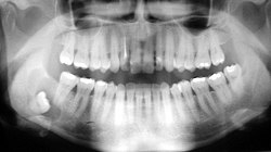
- Implantační: vzniká zatlačením epitelu do řídké pojivové tkáně v důsledku traumatu. Zatlačený epitel se začne množit a jeho vlastní výměšek časem vytvoří dutinu cysty. Příkladem jsou cysty vzniklé při pozánětlivých srůstech pobřišnice nebo zubní cysty.
- Hyperplastické: vzniká rozšířením žlázy nebo jejího vývodu. Je geneticky podmíněná a nejčastěji vzniká v mléčné žláze nebo v prostatě.
- Fetální: jedná se o vývojové anomálie a postihují různé orgány. Vznikají při nesprávném vývoji kůže, jater nebo ledvin nebo při persistenci některých emryonálních struktur.
- Nádorové: Některé nezhoubné i zhoubné nádory také vytvářejí cysty.
- Parazitární: S obsahem tekutiny a částí parazitů.
Nejznámější druhy cyst
- ovariální cysta (ve skutečnosti se jedná spíše o pseudocystu)
- zubní cysta
Odkazy
Literatura
- Prof. MVDr. Roman Halouzka, DrSc., et al. Obecná veterinární patologie. 1. vyd. v ČR. Brno: Veterinární a farmaceutická univerzita Brno, 2004, 188 s. ISBN 80-7305-496-5.
Související články
- Pseudotumor
- Pseudocysta
- Absces
- Polycystická choroba ledvin
Externí odkazy
 Obrázky, zvuky či videa k tématu cysta na Wikimedia Commons
  Slovníkové heslo cysta ve Wikislovníku
Přečtěte si prosím pokyny pro využití článků o zdravotnictví.
Média použitá na této stránce
Star of life, blue version. Represents the Rod of Asclepius, with a snake around it, on a 6-branch star shaped as the cross of 3 thick 3:1 rectangles.
Design:
The logo is basically unicolor, most often a slate or medium blue, but this design uses a slightly lighter shade of blue for the outer outline of the cross, and the outlines of the rod and of the snake. The background is transparent (but the star includes a small inner plain white outline). This makes this image usable and visible on any background, including blue. The light shade of color for the outlines makes the form more visible at smaller resolutions, so that the image can easily be used as an icon.
This SVG file was manually created to specify alignments, to use only integers at the core 192x192 size, to get smooth curves on connection points (without any angle), to make a perfect logo centered in a exact square, to use a more precise geometry for the star and to use slate blue color with slightly lighter outlines on the cross, the rod and snake.
Finally, the SVG file is clean and contains no unnecessary XML elements or attributes, CSS styles or transforms that are usually added silently by common SVG editors (like Sodipodi or Inkscape) and that just pollute the final document, so it just needs the core SVG elements for the rendering. This is why its file size is so small.ID#: 604 Description: Histopathology of Taenia saginata in appendix.
Histopathology of Taenia saginata in appendix. Parasite.
Content Providers(s): CDC Creation Date: 1971
Copyright Restrictions: None - This image is in the public domain and thus free of any copyright restrictions. As a matter of courtesy we request that the content provider be credited and notified in any public or private usage of this image.Autor: Uwe Gille, Licence: CC-BY-SA-3.0
opened uterus of a dog with a glandular-cystic hyperplasia of the endometrium
Peritoneal Cyst
This 8-centimeter cyst was incidentally found at hysterectomy floating free in the peritoneal cavity, a Zeppelin of the abdomen, if you will. Before making the diagnosis of this benign lesion, it is important for the pathologist to quiz the surgeon concerning the intra-abdominal findings. A cyst excised from a multicystic mesothelioma may look similar to this, and while multicystic mesothelioma may not be a true neoplasm, it has a stubborn tendency to recur locally.
The original photo was taken with a Nikon FE2, Nikkor macro lens, and Ektachrome Elite 100 film. The subject was lit by 4 photofloods. The film is balanced for daylight, so a blue compensation filter was used. The transparency was scanned with a Polaroid SprintScan 35 and edited with Photoshop 3.04. Digital editing included using the Dust and Scratches filter to remove lint from the background, and employing the Levels command to balance the dynamic range.
Photograph by Ed Uthman, MD. Public domain. Posted 30 May 99Retinierter Weisheitszahn mit zystischer Lyse im Unterkiefer.


