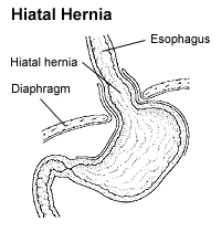
Hiátová hernie
| Hiátová hernie | |
|---|---|
 Hiátová hernie | |
| Klasifikace | |
| MKN-10 | K44. |
| Některá data mohou pocházet z datové položky. | |
Hiátová hernie (jícnová kýla[1], hiátová kýla, latinsky hiatus hernia) je vyklenutí horní části žaludku trhlinou v bránici nebo jícnovým otvorem do hrudníku. Vážnější formy se řeší chirurgickým zákrokem.[2][3]
Klasifikace hiátové hernie, jak ji provedl roku 1926 švédský rentgenolog Ake Akerlund dle 60 literárních a 30 vlastních pozorování.[4]
- Hiátová hernie se zkráceným jícnem, kde není možná repozice.
- Paraezofageální hiátová hernie.
- Ostatní hiátové hernie, kde jícen není zkrácen, ale distální konec ezofagu tvoří část obsahu kýlního vaku.
Příznaky
Nemusí být patrné, jinak např. bolest na hrudi, krvácení do zažívacího ústrojí.[2]
Odkazy
Reference
- ↑ Archivovaná kopie. www.iscare.cz [online]. [cit. 2015-09-26]. Dostupné v archivu pořízeném dne 2015-09-27.
- ↑ a b http://hiatova-kyla-hernie.nasclovek.cz/
- ↑ Archivovaná kopie. www.stefajir.cz [online]. [cit. 2014-12-30]. Dostupné v archivu pořízeném dne 2014-12-30.
- ↑ http://eportal.chirurgie.upol.cz/portal_final/?page_id=312
Literatura
- Akerlund A. I. Hernia diaphragmatica Hiatus oesophagei vom anatomischen und röntgenologischen Gesichtspunkt. Acta Radiol. 1926;6:3–22.
- Šimon J. Hiátová brániční kýla. Čas Lék Čes. 1928;67(12):423–427,462–466.
- Drahaňovský V. Refluxní nemoc jícnu a hiátové hernie. In: Duda M, Czudek S. Mininvazivní chirurgie. Třinec: Nemocnice Podlesí; 1996. s. 73–78.
- Duda M, Dlouhý M, Gryga A, Köcher M. Možnosti laparoskopických a torakoskopických operací v chirurgii jícnu a žaludku. In: Říha V, et al., editor. Endoskopická chirurgie. Sborník prací III. celostátní konference o laparoskopické chirurgii; 22.–23. dubna 1994; Benešov u Prahy.s. 74–79.
- Neoral Č, Král V. Laparoskopická fundoplikace. Rozhl Chir. 1996; 75(7):345–348.
- Neoral Č, Aujeský R, Král V. Místo antirefluxního výkonu v terapii refluxní nemoci jícnu – problém diagnostický, terapeutický a indikační. Čes a slov Gastroent. 1997;51(6):207–209.
Externí odkazy
 Obrázky, zvuky či videa k tématu hiátová hernie na Wikimedia Commons
 - Hiatus Hernia: rady a pomoc (anglicky)
- Denis P. Burkitt, M. D., D. Sc., F.R. C. S., FR. S: Hiatus hernia: is it preventable? (anglicky)
Přečtěte si prosím pokyny pro využití článků o zdravotnictví.
Média použitá na této stránce
Autor: James Heilman, MD, Licence: CC BY-SA 3.0
A large hiatus hernia as seen on CT
Star of life, blue version. Represents the Rod of Asclepius, with a snake around it, on a 6-branch star shaped as the cross of 3 thick 3:1 rectangles.
Design:
The logo is basically unicolor, most often a slate or medium blue, but this design uses a slightly lighter shade of blue for the outer outline of the cross, and the outlines of the rod and of the snake. The background is transparent (but the star includes a small inner plain white outline). This makes this image usable and visible on any background, including blue. The light shade of color for the outlines makes the form more visible at smaller resolutions, so that the image can easily be used as an icon.
This SVG file was manually created to specify alignments, to use only integers at the core 192x192 size, to get smooth curves on connection points (without any angle), to make a perfect logo centered in a exact square, to use a more precise geometry for the star and to use slate blue color with slightly lighter outlines on the cross, the rod and snake.
Finally, the SVG file is clean and contains no unnecessary XML elements or attributes, CSS styles or transforms that are usually added silently by common SVG editors (like Sodipodi or Inkscape) and that just pollute the final document, so it just needs the core SVG elements for the rendering. This is why its file size is so small.Author: National Institute of Diabetes and Digestive and Kidney Diseases, National Institutes of Health
Source URL: http://digestive.niddk.nih.gov/ddiseases/pubs/dictionary/pages/e-k.htm
Copyright tag: Why? Because it's from an NIH department.
Images


