Schistosoma
 Schistsoma mansoni | |
| Vědecká klasifikace | |
| Říše | živočichové (Animalia) |
| Kmen | ploštěnci (Platyhelminthes) |
| Třída | motolice (Trematoda) |
| Podtřída | Digenea |
| Řád | Strigeidida |
| Čeleď | Schistosomatidae (Krevničkovití) |
| Rod | Schistosoma Weinland, 1858 |
| druhy | |
S. mansoni | |
| Některá data mohou pocházet z datové položky. | |
Schistosoma (česky též krevnička, v anglické literatuře označované jako blood flukes) je medicínsky nejvýznamnější rod motolic na světě. Schistosomy jsou motolice odděleného pohlaví s výrazným pohlavním dimorfismem, které parazitují v cévní soustavě obratlovců, včetně člověka. Způsobují onemocnění člověka a zvířat - schistosomózu (dříve bilharzióza), které je známé už z dávných dob egyptských faraónů.
Morfologie
Schistosomy jsou na rozdíl od ostatních motolic odděleného pohlaví (gonochoristé) s výrazným pohlavním dimorfismem. Samičky jsou delší a štíhlejší než samci, na příčném řezu téměř kruhovitého tvaru a měří 10-20 mm x 0,1-0,3 mm.Samci měří 6-12 mm x 5-9 mm, jsou plochého tvaru těla, kaudálně od břišní přísavky se tegument stáčí a vytváří žlábek zvaný – canalis gynecophorus. V něm je v době kopulace uložená samička. Vajíčka schistosom jsou vřetenovitého až oválného tvaru, bez operkula, opatřené na jednom pólu trnem. Tento trn má jak patogenní tak diagnostický význam.
Vývojový cyklus
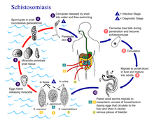
Dospělí jedinci schistosom se lokalizují v cévní soustavě savců a ptáků, nejčastěji v žilním řečišti střev, jater a močové soustavy. Samička je při kopulaci stočená v kanálku samce. Po kopulaci samička opouští kanálek samečka a migruje do cílových míst, kde klade vajíčka s typickými trny. Vajíčka pomocí hrotů a proteolytických enzymů provrtávají stěny kapilár a migrují tkáněmi do střeva (S. mansoni, S. japonicum) nebo do močového měchýře (S. haematobium). Vajíčka pak opouštějí definitivního hostitele trusem nebo močí a ve vodním prostředí se z vajíček líhnou obrvené larvy – miracidium. Miracidia aktivně plavou ve vodě a hledají vhodného mezihostitele, kterými jsou plži rodu Bulinus a Biomphalaria. Miracidium proniká do plže a mění se ve vakovitou mateřskou sporocystu. V ní se vyvíjí další dceřiné sporocysty. V těle dceřiných sporocyst poté vznikají cerkarie s vidličkovitým ocáskem – tzv. furkocerkarie. Ty plže opouští a plavou ve vodě. Do 72 hodin musí najít definitivního hostitele jinak hynou. Furkocerkarie hledají kůži hostitele na základě chemotaxe. Při kontaktu s kůží hostitele se do ní během 10 minut zavrtají a přemění se ve schistosomuly. Ty migrují následně pojivovými tkáněmi kůže a podkožím až do žil. Krevním oběhem jsou poté zaneseny do vrátnicové žíly, kde dospívají a kopulují.
Externí odkazy
 Obrázky, zvuky či videa k tématu Schistosoma na Wikimedia Commons
 - Ptačí schistosomy a cerkariová dermatitida
Média použitá na této stránce
Autor:
- Information-silk.png: Mark James
- derivative work: KSiOM(Talk)
A tiny blue 'i' information icon converted from the Silk icon set at famfamfam.com
ID#: 4841 Description: This micrograph depicts an egg from the parasite Schistosoma mansoni, and reveals the egg’s characteristic lateral spine.
S. mansoni eggs are large, measuring 114µm -180µm in length, and have a characteristic shape, with a prominent lateral spine near their posterior end. The anterior end is tapered, as well as slightly curved.
Content Providers(s): CDC Creation Date: 1979
Copyright Restrictions: None - This image is in the public domain and thus free of any copyright restrictions. As a matter of courtesy we request that the content provider be credited and notified in any public or private usage of this image.Title: Pathology: SEM: Schistosome Parasite
Description: This schistosome parasite enters the body through the skin of persons coming in contact with infested waters. The adult worm lives in the veins of its host. The parasite is magnified x256 in this photograph.
Subjects (names):
Topics/Categories: Pathology -- Scanning Electron Microscopy (SEM)
Type: Black & White Print. Black & White Slide
Source: National Cancer Institute
Author: Bruce Wetzel (photographer). Harry Schaefer (phot
AV Number: AV-0000-3692
Date Created: Unknown
Date Entered: 1/1/2001
Access: PublicEggs are eliminated with feces or urine (1). Under optimal conditions the eggs hatch and release miracidia (2), which swim and penetrate specific snail intermediate hosts (3). The stages in the snail include 2 generations of sporocysts (4) and the production of cercariae (5). Upon release from the snail, the infective cercariae swim, penetrate the skin of the human host (6), and shed their forked tail, becoming schistosomulae (7). The schistosomulae migrate through several tissues and stages to their residence in the veins (8,9). Adult worms in humans reside in the mesenteric venules in various locations, which at times seem to be specific for each species (10). For instance, S. japonicum is more frequently found in the superior mesenteric veins draining the small intestine [A], and S. mansoni occurs more often in the superior mesenteric veins draining the large intestine [B]. However, both species can occupy either location, and they are capable of moving between sites, so it is not possible to state unequivocally that one species only occurs in one location. S. haematobium most often occurs in the venous plexus of bladder [C], but it can also be found in the rectal venules. The females (size 7 to 20 mm; males slightly smaller) deposit eggs in the small venules of the portal and perivesical systems. The eggs are moved progressively toward the lumen of the intestine (S. mansoni and S. japonicum) and of the bladder and ureters (S. haematobium), and are eliminated with feces or urine, respectively (1).


