Vnitřní fixace

Vnitřní fixace (interní fixace) je chirurgický úkon, který se obvykle (na rozdíl od zevní fixace) používá při operativní léčbě (osteosyntéze) „méně“ komplikovaných zlomenin kostí. Jde o ortopedický/traumatologický operační přístup, při kterém je celý osteosyntetický implantát (hřeb, dlaha, šroub, drát nebo svazek drátů, cerkláž atp.) kryt měkkými tkáněmi nebo uložen na povrchu či uvnitř kosti. Po ukončení léčby zůstává implantát ve většině případů trvale uvnitř těla.
Vnitřní fixace je také stále důležitým tématem v biomechanice.
Operativní přístupy vnitřní fixace
Existuji dva hlavní přístupy k vnitřní fixaci:
- ORIF (anglicky Open Reduction Internal Fixation, česky otevřená repozice a vnitřní fixace).
- CRIF (anglicky Closed Reduction Internal Fixation, česky uzavřená repozice a vnitřní fixace).
V mnoha případech se lékařské indikace pro vnitřní fixaci překrývají s indikací pro zevní fixaci.
Materiály používané při vnitřní fixaci
Hlavním požadavkem na materiály používané při vnitřní fixaci je biokompatibilita.
Běžně používané materiály implantátů patří především chirurgická nerezová ocel a chirurgické titanové slitiny. Dále se používají slitiny kobaltu a chrómu, čistý titan nebo plasty aj.
Zvláštní pozornost je také věnována kvalitě a úpravě povrchu implantátů (např. anodizace atp.).
Historie vnitřní fixace
Koncept vnitřní fixace pochází z poloviny 19. století. Vnitřní fixace se stala "rutinní" záležitostí v polovině 20. století.
Na začátku 20. století významně ovlivnil rozvoj vnitřní fixace belgický chirurg Albin Lambotte, používal různé druhy vnitřní osteosyntézy, od cerkláží přes šrouby až po dlahy.
Významným mezníkem bylo založení švýcarské společosti AO (Arbeitsgemeinschaft für Osteosynthesefragen, česky Společnost pro otázky osteosyntézy) v roce 1958.
Příklady aplikace vnitřní fixace

Zlomenina radia a ulny a její léčení vnitřní fixací (dlahy).
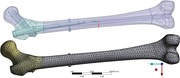
Lidský femur se zlomeninami AO 31B3 (fractura mediocervicalis femoris) a AO 31A3 (fractura subtrochanterica femoris) a s vloženým intramedulárním hřebem Tantum (a) CAD model, (b) zjednodušená síť konečných prvků pro výpočet pomocí metody konečných prvků.[2]
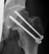
RTG snímek − Osteosyntéza krčku femuru člověka pomocí kovových šroubů.
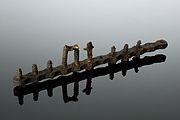
Kostní dlaha, Evropa z let 1914−1918.
Reference
Související články
Externí odkazy
 Obrázky, zvuky či videa k tématu vnitřní fixace na Wikimedia Commons

Přečtěte si prosím pokyny pro využití článků o zdravotnictví.
Média použitá na této stránce

Star of life, blue version. Represents the Rod of Asclepius, with a snake around it, on a 6-branch star shaped as the cross of 3 thick 3:1 rectangles.
Design:
The logo is basically unicolor, most often a slate or medium blue, but this design uses a slightly lighter shade of blue for the outer outline of the cross, and the outlines of the rod and of the snake. The background is transparent (but the star includes a small inner plain white outline). This makes this image usable and visible on any background, including blue. The light shade of color for the outlines makes the form more visible at smaller resolutions, so that the image can easily be used as an icon.
This SVG file was manually created to specify alignments, to use only integers at the core 192x192 size, to get smooth curves on connection points (without any angle), to make a perfect logo centered in a exact square, to use a more precise geometry for the star and to use slate blue color with slightly lighter outlines on the cross, the rod and snake.
Finally, the SVG file is clean and contains no unnecessary XML elements or attributes, CSS styles or transforms that are usually added silently by common SVG editors (like Sodipodi or Inkscape) and that just pollute the final document, so it just needs the core SVG elements for the rendering. This is why its file size is so small.
Autor: Fry72 Karel Frydrýšek, Tomáš Halo, Daniel Čepica, Licence: CC BY-SA 4.0
Biomechanika, Femur s hřebem, CAD model, síť konečných prvků pro metodu konečných prvků
Autor: Fry72 Karel Frydrýšek, Licence: CC BY-SA 4.0
Příklad využití vnitřní fixace při osteosyntéze (rtg. snímek - subtrochanterická liniová zlomenina femuru s aplikací intramedulárního hřebu v umělé kosti resp. subtrochanterická zlomenina s interfragmentem resp. tříštivá zlomenina proximálního femuru).

taken by myself available online at http://www.bakedbabies.com/comics/images/strip167.png
i am the creator of this original work. i am licensing this file as public domain

Autor: unknown, Licence: CC BY 4.0
Bone plates are used to support fractures and encourage them to heal in proper alignment. This rather rusty example of twelve screws fixed into a plate is believed to have been put in position by Sir William Arbuthnot Lane (1856–1943), a leading surgeon and health campaigner.
Lane pioneered the technique of internal fixing of fractures as opposed to using external splints. Lane inserted his first internal splint in 1894, and after 1905 he introduced his steel bone plates and screws. His methods were soon taken up in the United States and Germany but not in Britain until 1912.
maker: Unknown maker
Place made: Europe
Wellcome Images
Keywords: orthopaedics; bone plate

Autor: Fry72 Karel Frydrýšek, Milan Šír, Leopold Pleva, Licence: CC BY-SA 4.0
Femur, Zlomenina, Collum femoris, Lékařské šrouby (vruty), podložky, RTG