Vysokohorský otok plic
Vysokohorský otok plic (zkratka VOP) je vážnou formou akutní horské nemoci, i když tato onemocnění spolu nemusí přímo souviset.
Léčba je podobná jako u vysokohorského otoku mozku, je nutný rychlý sestup do nižší nadmořské výšky, při zanedbání tohoto faktu hrozí postiženému smrt. Po sestupu se organismus většinou sám zregeneruje do 2 dnů, po vymizení příznaků je možné pokračovat ve výstupu.
Typické příznaky VOP
- extrémní únava
- nemožnost popadnout dech
- modré či šedé rty, případně nehty
- chrčivé nebo bublavé dýchání
- kašel
- sevření nebo tlak v prsou
Externí odkazy
Přečtěte si prosím pokyny pro využití článků o zdravotnictví.
Média použitá na této stránce
Star of life, blue version. Represents the Rod of Asclepius, with a snake around it, on a 6-branch star shaped as the cross of 3 thick 3:1 rectangles.
Design:
The logo is basically unicolor, most often a slate or medium blue, but this design uses a slightly lighter shade of blue for the outer outline of the cross, and the outlines of the rod and of the snake. The background is transparent (but the star includes a small inner plain white outline). This makes this image usable and visible on any background, including blue. The light shade of color for the outlines makes the form more visible at smaller resolutions, so that the image can easily be used as an icon.
This SVG file was manually created to specify alignments, to use only integers at the core 192x192 size, to get smooth curves on connection points (without any angle), to make a perfect logo centered in a exact square, to use a more precise geometry for the star and to use slate blue color with slightly lighter outlines on the cross, the rod and snake.
Finally, the SVG file is clean and contains no unnecessary XML elements or attributes, CSS styles or transforms that are usually added silently by common SVG editors (like Sodipodi or Inkscape) and that just pollute the final document, so it just needs the core SVG elements for the rendering. This is why its file size is so small.Autor: Maryrosegrant, Licence: CC BY-SA 4.0
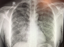
Plain chest x-ray (radiograph) of a patient diagnosed with HAPE. There are patchy infiltrates throughout the lung tissue, with predominant changes in the right middle lobe/right central hemithorax.


