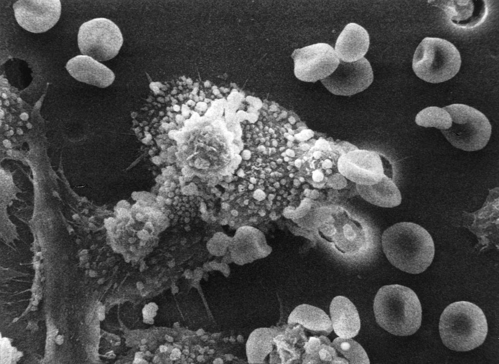
Macs killing cancer cell
Relevantní obrázky
Relevantní články
Biologická léčba nádorových onemocněníBiologickou léčbou nádorových onemocnění nazýváme takový způsob léčby, který k odstranění nádorů využívá prvky a procesy imunitního systému zasaženého organismu. K takové léčbě řadíme například aktivní imunizaci proti nádorovým buňkám pomocí speciálního očkování, kdy jsou vlastní leukocyty nemocného obohaceny o rozpoznávací systém, který je typický pro daný nádor. Na obdobném principů funguje aktivace výkonných imunocytů, a to buď pomocí cytokinů, nebo odběrem těchto imunocytů, jejich následným propojením s protilátkou proti antigenu nádoru, a vrácením za účelem jejich boje proti nádoru. Rozšířením takové imunoterapie je obohacení těchto specifických monoklonálních protilátek buď o chemoterapeutické (cytostatické) léčivo, nebo radioaktivní izotop. Po podání takto obohacených protilátek dojde k tomu, že protilátky "vyhledají" nádorovou tkáň a zároveň k ní "přinesou" příslušné protinádorové léčivo. .. pokračovat ve čtení
ImunoterapieBiologická léčba nebo imunoterapie, řídč. cílená léčba, využívá imunitu organismu a její zeslabení nebo zesílení k léčbě nemocí. Imunoefektorové buňky typu lymfocytů, makrofágů, dendritických buněk, NK buňky, T-lymfocyty mohou účinně bojovat proti rakovině či některým autoimunitním chorobám díky tomu, že jsou lépe známé struktury a pochody na povrchu i uvnitř buňky. Terapie granulocyte colony-stimulating factor (G-CSF), interferonů, imikvimodů a buněčné membránové frakce bakterií jsou povoleny pro medicínské použití. Další typy terapií IL-2, IL-7, IL-12, chemokiny, (CpG) oligodeoxynukleotidy a glukany procházejí klinickým hodnocením. .. pokračovat ve čtení
MakrofágMakrofág je buňka přirozené imunity, která hraje velmi důležitou roli v imunitní reakci. Jedná se o zástupce mononukleárů, tj. buněk s jedním, nesegmentovaným jádrem. .. pokračovat ve čtení


