Picomonas judraskeda
Autor:
Ramkumar Seenivasan, Nicole Sausen, Linda K. Medlin, Michael Melkonian
Přisuzování:
Obrázek je označen jako „Vyžadováno uvedení zdroje“ (Attribution Required), ale nebyly uvedeny žádné informace o přiřazení. Při použití šablony MediaWiki pro licence CC-BY byl pravděpodobně parametr atribuce vynechán. Autoři zde mohou najít příklad pro správné použití šablon.
Shortlink:
Zdroj:
Formát:
3098 x 4217 Pixel (10183378 Bytes)
Popis:
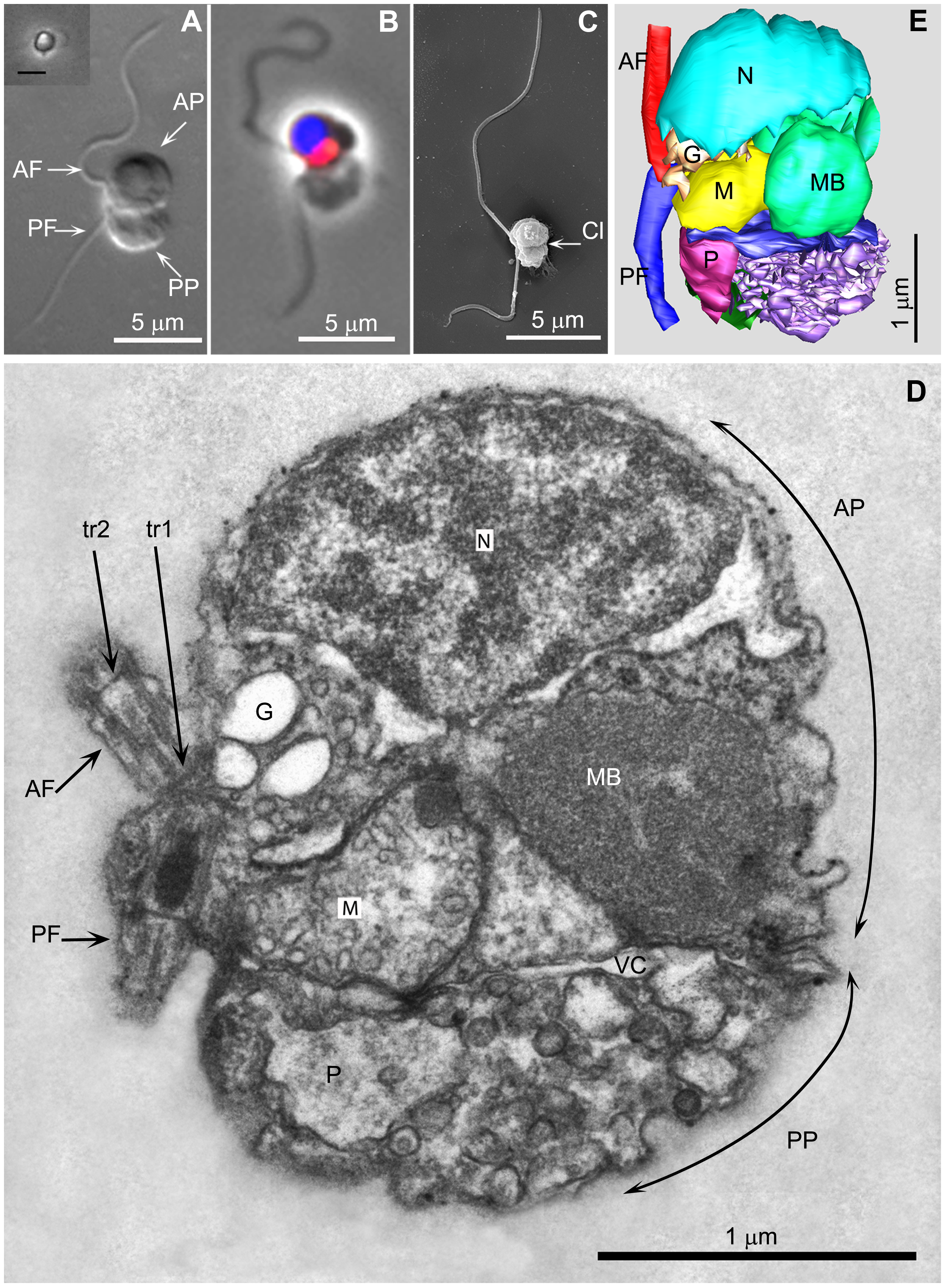
A Picomonas cell.
- A. Differential interference contrast of a chemically fixed cell. Inset shows phase contrast image of a live cell from tissue culture flask photographed with an inverted microscope (Scale bar 5 µm).
- B. Fluorescence and phase contrast overlay, nucleus (blue), mitochondrion (red).
- C. SEM image.
- D. A longitudinal section through a cell in the plane of the flagella, viewed from the cell’s left.
- E. A 3 D serial section reconstruction of the cell depicted in 2D. AF/PF (anterior−/posterior flagellum); AP/PP (anterior/posterior part of the cell); G (Golgi body); M (mitochondrion); MB (‘microbody’); N (nucleus); tr1,tr2 (distal [tr2] and proximal [tr1] flagellar transitional regions); P (posterior digestive body); Cl (cleft separating the anterior from the posterior part of the cell); vc (vacuolar cisterna).
Komentář k Licence:
Toto dílo bylo publikováno v rámci Veřejné vědecké knihovny. Její webová stránka prohlašuje, že obsah všech PLOS periodik je publikován pod licencí Creative Commons Attribution 2.5.
Pro nahrávání: Musíte poskytnout odkaz (URL) k originálnímu souboru nebo článku v periodiku.
Licence:
Credit:
Sdílet obrázek:


Relevantní články
PicozoaPicozoa je skupina mořských jednobuněčných pikoplanktonních organismů, které jsou s největší pravděpodobností příbuzné ruduchám. Od r. 2007 byl znám z genomové analýzy environmentálních vzorků a nazýván Picobiliphyta, ale teprve v r. 2013 se podařilo kultivovat jednoho ze zástupců, Picomonas judraskeda. Objev způsobil zásadní změnu představ o této skupině, zpochybnil fototrofní způsob obživy a vedl tak i k přejmenování na současný název. .. pokračovat ve čtení