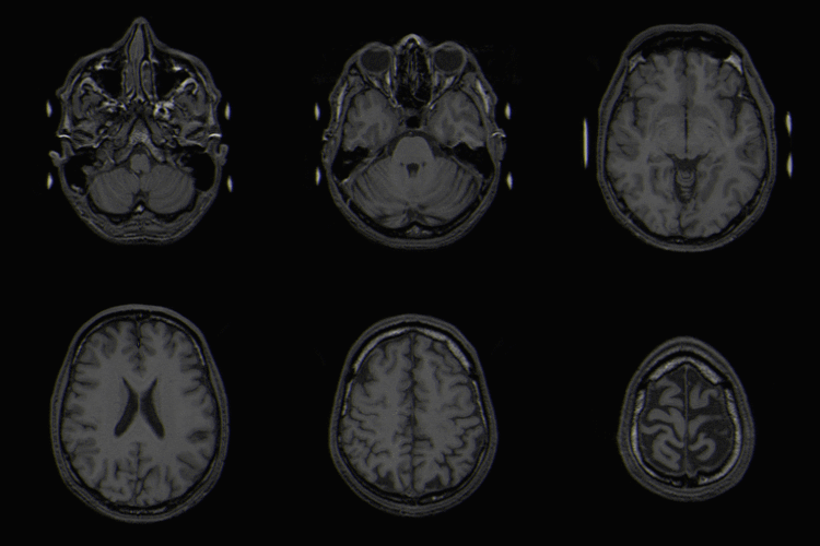
Melanoma met ax t2
Autor:
Dr Laughlin Dawes
Přisuzování:
Obrázek je označen jako „Vyžadováno uvedení zdroje“ (Attribution Required), ale nebyly uvedeny žádné informace o přiřazení. Při použití šablony MediaWiki pro licence CC-BY byl pravděpodobně parametr atribuce vynechán. Autoři zde mohou najít příklad pro správné použití šablon.
Shortlink:
Zdroj:
Formát:
698 x 759 Pixel (84756 Bytes)
Popis:
"This 59 year-old female patient presented with acute right hemiplegia, aphasia and confusion. She had a known cerebral melanoma metastasis in the left frontal lobe. This axial T2-weighted MR (click image for arrows) shows a large haematoma with a fluid-fluid level (green arrows). There is a smaller low signal area anteriorly (red arrows) which corresponded to the known metastasis. This smaller area enhanced after gadolinium, as did the overlying dura." (dr Dawes)
Komentář k Licence:
author kindly mailed me permission to use this and other images on cc-by-3.0 license
Licence:
Credit:
Relevantní obrázky
Relevantní články
NeuroonkologieNeuroonkologie je specializace lékařství, která spojuje neurologii a onkologii. .. pokračovat ve čtení


