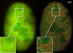
TMV virus super resolution microscopy Christoph Cremer Christina Wege
B: after Gaussian blurring corresponding to the individual localization accuracy D: same data, but distribution of the individual molecule position without Gaussain blurring
Virus Super Resolution Microscopy/Optical nanoscopy: A further example for the use of localization microscopy/SPDM is an analysis of Tobacco mosaic virus (TMV) particles. TMV particles are not only important in plant research but are also an example for a class of most promising biomolecule complexes exhibiting a high potential for nanotechnology applications as virus-derived biotemplates for nanostructured hybrid materials and building-blocks in analytical, technical & therapeutic applications.
Collaboration of the former Christoph Cremer lab, emeritus at Heidelberg university [1], [2] & the Wege lab (Universities of Stuttgart).
Reference: Superresolution imaging of biological nanostructures by spectral precision distance microscopy (2011): Cremer, R. Kaufmann, M. Gunkel, S. Pres, Y. Weiland, P- Müller, T. Ruckelshausen, P. Lemmer, F. Geiger, S. Degenhard, C. Wege, N- A. W. Lemmermann, R. Holtappels, H. Strickfaden, M. Hausmann , Biotechnology 6: 1037–1051Relevantní obrázky
Relevantní články
Superrozlišovací mikroskopieSuperrozlišovací mikroskopie je optická mikroskopie umožňující pozorovat objekty s rozlišením vyšším než difrakční limit. Ten při běžné světelné mikroskopii neumožňuje odlišit dva body bližší než přibližně 250 nm Za pokroky ve vývoji těchto metod v roce 2014 získali Eric Betzig, Stefan W. Hell a William E. Moerner Nobelovu cena za chemii. .. pokračovat ve čtení


